42 chromosome diagram unlabeled
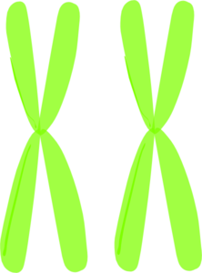
Learn the parts of a cell with diagrams and cell quizzes - Kenhub For this exercise we'll start with an image of a cell diagram ready labeled. Study this and make sure that you're clear about which structure is found where. Cell diagram unlabeled It's time to label the cell yourself! As you fill in the cell structure worksheet, remember the functions of each part of the cell that you learned in the video. Anita thinks that she has heterozygous alleles for red hair. If she is ... If she is correct, which of these diagrams best illustrates Anita s alleles for red hair? 2 chromosomes are labeled Upper R and Upper R. 2 chromosomes are labeled Upper R and r. One chromosome is labeled Upper R and another is unlabeled. 2 chromosomes are labeled r and r. Heterozygous is where the alleles of diploid organisms are different.
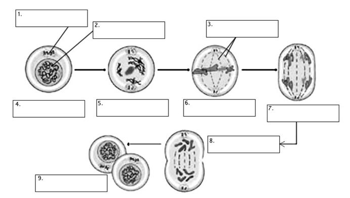
Animal Cell Telophase Diagram / Cell division...Prophase, Prometaphase ... Unlabeled animal cell diagram finally an unlabeled version of the diagram is included at the bottom of the cytokinesis drawing animal cell diagram telophase and image information: Nuclei are forming around the 4 groups of condensed. Simplified diagram of an animal cell.
Chromosome diagram unlabeled
Dna Chemical Structure Diagram - Drawing Diagram These instructions are stored inside each of your cells distributed among 46 long structures called chromosomes. Download scientific diagram Chemical structure of DNA. Size of this PNG preview of this SVG file. Zooming in on DNA Structure. DNA is the information molecule. Base Pair Of Nucleic Acids Chemistry Molecular Biology Chemical Structure Somatic chromosomes of the grasses Zingeria trichopoda and Zingeria ... However, these interstitial signals are due to the incomplete blocking by the unlabeled DNA and, therefore, not indica- tive of an intergenomic translocation. Results from 457 interphase nuclei... Bacteria in Microbiology - shapes, structure and diagram Bacterial spores. Bacterial endospores layers. Bacteria cells are the smallest living cells that are known; even though viruses are smaller than bacteria, viruses are not living cells. There are different types of bacteria with various sizes, shapes, and structures. The bacteria shapes, structure, and labeled diagrams are discussed below.
Chromosome diagram unlabeled. Animal Cell Telophase Diagram : The Diagram Given Below Represents A ... Learners need to know the names of the phases and they need to be able to draw simple descriptive diagrams showing the chromosome changes. ... Unlabeled animal cell diagram finally an unlabeled version of the diagram is included at the bottom of the cytokinesis drawing animal cell diagram telophase and image information: Source: d3i71xaburhd42 ... Imaging unlabeled proteins on DNA with super-resolution As a first demonstration of the ability to visualize unlabeled proteins on DNA, we studied human recombinase protein RAD51 (hRAD51) using DNA-intercalation-based inverse imaging. Here, DNA was incubated with hRAD51 in a calcium-containing buffer to form DNA-bound hRAD51 filaments ( 27, 28) (Materials and Methods). Unlabelled Plant Cell Diagram Gcse - Blogger Diagrams from past papers identify, labelled and unlabelled parts and give the function of parts. Human cell diagram parts pictures structure and functions. Running through the petiole are vascular bundles, which then form the veins in the leaf. Cytoplasm is found inside the cell and contains all the other cell structures. Figure 13.4b shows an autoradiograph of a replicating bact ... - Chegg FIGURE 13.4 The process of bacterial chromosome replication. (b) A replicating E. coli chromosome visualized by autoradiography and transmission electron microscopy (TEM). This chromosome was radiolabeled by growing bacterial cells in media containing radiolabeled thymidine. The diagram at the right shows the locations of the two replication forks.
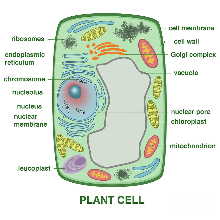
Structure of Chromosomes (With Diagram) | Cell Nucleus | Biology The gene is a part of the DNA molecule. ADVERTISEMENTS: Each chromosome consists of one to four coiled threads called chromonema and also contains juxtaposed minute particles known as chromomeres (Fig. 1.15) which are rich in DNA. Most of the chromosomes possess usually two constrictions - primary (kinetochore) and secondary. Unlabeled Clip Art - Royalty Free - GoGraph Download high quality Unlabeled clip art from our collection of 66,000,000 clip art graphics. 800-810-1617 gograph@gograph.com ... Medical Labeled Diagram Closeup With Muscle, Transverse Carpal Ligament, Median Nerve, Tendon Sheath, Flextor Tendons And Bones. ... Educational And Medical Scheme With Cell, Chromosome And Dna. Labeled Anatomical ... Blank Diagram Of A Dna Structure - Drawing Diagram Blank diagram of a dna structure.April 11th 2019 - DNA Structure By Cindy Grigg organisms structure valuable newly enzyme evidence alike nucleus dominant unless radiation mutation organism enzymes ferret structures synthesis rung Directions Fill in each blank with the word that best completes the reading comprehension An embryonic cell divides again and again Where there was one cell NAME. A Labeled Diagram of the Plant Cell and Functions of its Organelles The nucleus is known to be the 'control room' of the cell. It regulates various cell functions by controlling the protein synthesis of the plant cell. The nucleus contains DNA within the chromosomes. It is a membrane-bound structure that contains the cells hereditary information. Function: Controls expression and transcription of the gene ...
Chromosome constitutions of SHW-L1/rye lines. In a, c, d, e, and g ... Download scientific diagram | Chromosome constitutions of SHW-L1/rye lines. ... (yellow), and pAs1 (red) probes, with unlabeled wheat genome DNA used as a blocker. a Line 186-14-18-8-8-27 had 28A ... Chromosome Clipart Teaching Resources | Teachers Pay Teachers 2. $3.00. PDF. Google Apps™. These interactive diagrams are terrific for science teachers teaching their students about Chromosome and Chromosome Structure in a distance learning or blended learning environment. These click-and-learn diagrams are Google Slides files designed to be used in several possible ways.*. A Labelled Diagram Of Meiosis with Detailed Explanation The diagram of meiosis is beneficial for class 10 and 12 and is frequently asked in the examinations. The diagram of meiosis along with the explanation of its different stages is given below in detail. Further Reading: Meiosis II. Significance of Meiosis. Meiosis I. Well-Labelled Diagram for Meiosis. Meiosis I Prophase I. Here, the chromosomes ... Draw And Label Diagrams Of Plant Cell And Animal Cell : Printable ... Cells form the basic building blocks for all living things. Centrioles help move chromosomes during cell division. A comparison of plant and animal cells using labelled diagrams and descriptive explanations. Label the plant cell diagram using the glossary of plant cell terms. A system of flattened membranes called cisternae (mainpoint:
Cell Cycle (notes: 9.1)
Genetics Final (Multiple Choice) Flashcards - Quizlet Suppose that individual who carries the chromosomes in the diagram above is Aa. If A is on 3, then _ is on 4, and _ is on 5. ... What proportion of DNA molecules will be labeled with radioactivity after two rounds of replication in the presence of unlabeled nucleotides? 1/2. Which process would be most directly affected by deleting a promoter?

Free Prokaryote Cliparts, Download Free Prokaryote Cliparts png images ...
Solved Transcribed image text: Given the diagram below, assume that a G1 chromosome (e underwent one round of replication in thymidine and the metaphase chromosome (right) had both chromatids labeled. Which of the following replicative models conservative depressive seniservative could be eliminated by this observation?

33 Label A Bacterial Cell - Labels For You
PDF Mitosis Diagram Unlabeled april 18th, 2019 - g1 phase the cell grows s phase the cell makes copies of its chromosomes each chromosome now consists of two sister chromatids g2 phase the cell checks the duplicated chromosomes and gets ready to divide m phase the cell separates the copied chromosomes to form two full sets mitosis and cell diagram worksheet homeschooldressage …
December - Mrs. Beltz's Biology Class
PDF Lysosomes Diagram Labeled Pdf Download Heart Diagram Labeled DrawingCreate A Tube From The Top Of The Right Atrium, To Create The Superior Vena Cava. Let The Tube Fork Be Almost The Same Length As The Bump You Made To The Atrium's Right Chamber; Draw The Tube Of The Rounded Aorta Parallels The Bump.

Meiosis I | Biology for Non-Majors I
Diagram Lists - Stevenson High School You may wish consider printing the unlabeled diagrams, and labeling the significant structures yourself as you read or as diagrams are discussed in class. As an AP student, you are expected to be an active participant, so bring all diagrams to class for every unit. ... 12.4: Chromosome distribution and duplication during cell division 12.5: The ...
Homologous Chromosomes Clip Art at Clker.com - vector clip art online ...
DOC Mitosis: Labeled Diagram - West Branch High School The chromosomes line up separately on the spindle for metaphase of mitosis, but at the first division of meiosis the chromosomes pair in a process called synapsis. In synapsis, homologous chromosomes pair tightly along their length and then move to opposite poles so that only one of each pair of chromosomes ends up in each cell after the first ...
Plant Cell Photograph by Gwen Shockey
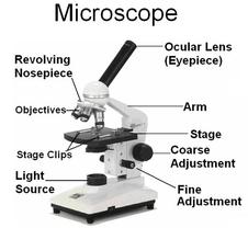
Diagram of Chromosome Structure - Online Biology Dictionary The name chromosome, meaning "colored body," is derived from the fact that in early studies of cellular structure the chromosomes could be easily stained with colored dyes and therefore showed up as colored bodies under the microscope. The diagram of chromosome structure above shows how DNA is organized in a eukaryotic cell.

Alila Medical Media | Cell, Molecular Biology & Genetics Images
Animal Cell Diagram Labeled Icev / Plant Cells Vs Animal Cells With ... Printable Animal Cell Diagram Labeled Unlabeled And Blank from i2.wp.com Separates chromosomes and is found as paired structures near the nucleus. The well labelled diagram of an animal cell consists of all the organelles and the structural components of an animal cell. Animal cell diagram for kids labeled.

Videos, Links, Extra Practice - Welcome to Ms. Thompson's Class Website
Lysosomes Diagram Labeled Pdf Download Heart Diagram Labeled DrawingCreate A Tube From The Top Of The Right Atrium, To Create The Superior Vena Cava. Let The Tube Fork Be Almost The Same Length As The Bump You Made To The Atrium's Right Chamber; Draw The Tube Of The Rounded Aorta Parallels The Bump.
Post a Comment for "42 chromosome diagram unlabeled"